Lab Reagents for Collaborations

Decorative photo Sandra Orsulic UCLA

Genetically Defined Mouse Ovarian Cancer Cell Lines

Decorative photo Sandra Orsulic UCLA mouse ovarian cancer models

Syngeneic Immunocompetent Mouse Ovarian Cancer Models

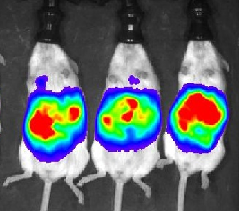
Orthotopic inoculation of syngeneic mouse ovarian cancer cell lines into FVB mice (PDF link) recapitulates key aspects of human ovarian cancer pathophysiology. These models are suitable for preclinical testing of drugs that may interfere with the immune system or testing the roles of the immune system in ovarian cancer progression.
Tissue Microarray Decorative photo Sandra Orsulic UCLA

Ovarian Cancer Tissue Microarrays (TMA)

  1. TMA of matched primary, metastatic, and recurrent samples from 42 patients (triplicate 1 mm cores for each of the primary high-grade serous ovarian cancer (HGSOC), concurrent pre-treatment metastasis, and recurrent post-treatment metastasis). The TMA is accompanied by detailed clinical data and an image database of immunohistochemical stains for markers of different immune cell types and fibroblasts. This TMA is useful for spatiotemporal studies of cell types involved in ovarian cancer progression. Contact: Sandra Orsulic, PhD
  2. TMA of serous (n=30), clear cell (n=28), endometrioid (n=18), mucinous (n=9), and transitional (n=9) ovarian cancer (duplicate 1mm cores). This TMA is useful for studies of different histologic subtypes of ovarian cancer. Contact: Sandra Orsulic, PhD
Decorative photo Sandra Orsulic UCLA

Expression Datasets

  1. NanoString data (https://www.ncbi.nlm.nih.gov/geo/query/acc.cgi?acc=GSE135712). Omental metastasis samples from 152 patients with HGSOC. The dataset is accompanied by detailed clinical data and an H&E image database with quantified content of fibroblasts, immune cells, and cancer cells. This resource supplements the publicly available expression datasets, which were primarily obtained from primary HGSOC.
  2. RNA-seq data (https://www.ncbi.nlm.nih.gov/geo/query/acc.cgi?acc=GSE138866). Omental metastasis samples from 130 patients with HGSOC. The dataset is accompanied by detailed clinical data and an H&E image database with quantified content of fibroblasts, immune cells, and cancer cells. This resource supplements the publicly available expression datasets, which were primarily obtained from primary HGSOC.
  3. RNA-seq data (https://www.ncbi.nlm.nih.gov/geo/query/acc.cgi?acc=GSE133296). Matched primary tumor, omental metastasis, and non-omental metastasis from 10 HGSOC patients. The dataset is accompanied by detailed clinical data and an H&E image database with quantified content of fibroblasts, immune cells, and cancer cells. This resource is useful for analysis of the tumor microenvironment at different physical locations in the peritoneal cavity.
  4. RNA-seq data and image database of normal fallopian tubes from 74 patients of different ages, menopausal status, and BRCA status (Forthcoming).
Photo of laboratory test tubes Orsulic Lab UCLA

Plasmids

RCAS plasmids (myr-Akt, myc, K-ras) can be obtained from Addgene http://www.addgene.org/pgvec1 (search Orsulic).